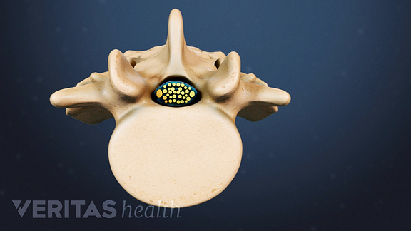
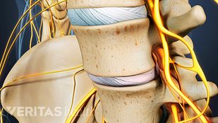
在脊髓组织中的退行性变化,在绝大多数的腰椎管狭窄症的诊断的老化结果相关联。1,2一个comprehensive evaluation of the potential sources is important for an accurate diagnosis of this condition.
椎管狭窄引起的脊髓神经的空间的收缩。看:腰椎管狭窄视频
Common Causes of Lumbar Spinal Stenosis
老化相关的变性可影响脊柱关节,韧带和/或椎间盘,引起以下变化2:
Hypertrophy of facet joints
变性关节炎可能导致关节面成为关节炎及生长骨刺(骨质长满),可能对脊神经根和/或脊髓的冲击。
看到Bone Spurs (Osteophytes) and Back Pain
Hypertrophy of ligamentum flavum
韧带的短带连接椎管内表面的增厚可压迫脊髓。
光盘的退化
Degeneration can cause a decrease in the height of intervertebral discs, reducing the disc space and narrowing the bony openings for spinal nerves (intervertebral foramen). Degeneration may also cause碟片疝,压缩脊神经根,和/或脊髓。
Degenerative spondylolisthesis
在椎骨中年龄相关的变化也可以在运动节段内引起不稳定,从而导致在其下方的一个椎体的打滑。
看到滑脱
一个条件可能会导致其他的,例如,在盘高度的降低可能导致膨胀或黄韧带屈曲进入椎管的背面。
不太常见的原因腰椎管狭窄症
很少,下面的情况可能会导致在后腰椎管狭窄:雷竞技下载网址
- Inherited or genetic,如短椎弓根(即在后面在前面连接椎体的拱椎骨的一部分)2
- Complications after lumbar surgery,如椎板切除术要么lumbar fusion2
- Systemic conditions,如佩吉特氏病2
- Spinal growths,如肿瘤,囊肿,或脓肿在下部脊柱(脓的集合)
腰椎管狭窄症的风险通常随着年龄的增加而增加。风险因素还包括遗传倾向,体重超标,涉及重体力劳动的职业。1
腰椎管狭窄症的诊断
一个doctor can diagnose stenosis in the lumbar spine based on specific clinical presentations and through medical imaging tests. Conducting a physical exam and reviewing the medical history helps a doctor determine the pattern of symptoms. Medical imaging tests help determine the location and severity of stenosis.
Physical exam and review of medical history
医生通常会检查疼痛、麻木、音乐le reflexes, and nerve function in the legs. Specific elements of the physical exam will include one or more of the following:
- 容安澜的测试。此测试检查脊髓损伤。在这个测试中,患者站立不支持,并与他们闭着眼睛。平衡的损失表明阳性测试结果,并且可以用信号L4-L5和/或L5-S1之间脊髓损伤或严重腰神经根。3
- 步态测试。In this test, the doctor analyzes the walking pattern of the patient to check for a wide-based or steppage gait, or loss of balance while walking.
- 神经系统检查。These tests include analyzing the leg’s muscle reflex to check for nerve root compression in the lower back.
- Straight leg raise test.这个测试的目的在于获得坐骨神经根压迫在腰椎和/或骶棘。在试验过程中,在他们的后面,医生轻轻患者躺在提升病人的腿。如果疼痛是这个动作过程中经历的测试被认为是积极的。
在一次体检中,医生还回顾病史,包括有关的发病和症状,既往手术,药物持续时间,以及随之而来的医疗条件存在的信息。雷竞技下载网址
医学影像检查为腰椎管狭窄症
影像学检查可能有助于定位腰椎椎管狭窄的部位和严重程度包括:
- 磁共振成像(MRI)。MRI检查通常被认为是黄金标准测试,以确定椎管狭窄。MRI的在识别的大小,形状,和脊柱组织之间的关联敏感,并且可以提供重要的结构,诸如退化的盘的信息。4,五
- X光片。常规的x射线在识别骨折和椎骨的缺陷,则运动节段的对准,椎间盘高度损失和骨骨刺形成是有用的。4
- 计算机断层扫描(CT)扫描。一个CT scan提供了脊柱结构的可视化,并且当MRI不可用,或者可能可以进行。
- 脊髓造影:一个脊髓造影involves the injection of a radiographic dye into the spinal canal, which is followed by a CT scan. Myelograms are typically only performed in patients who cannot undergo an MRI or have had prior surgery.
It is important to understand that medical image findings may not always correlate with the symptoms. Up to 30% of adults older than 60 years may have imaging evidence of lumbar spinal stenosis with no symptoms.2 A history of clinical symptoms along with imaging evidence is essential to diagnose symptomatic lumbar spinal stenosis.4
一旦腰椎管狭窄症被识别为患者的症状的原因,一个明确的治疗方案可配制。非手术治疗通常推荐了好几个星期被认为是外科手术之前。
参考
- 1。Huang W, Zhou G, Zhang Y. Risk factors for ligamentum flavum hypertrophy in lumbar spinal stenosis patients from the Xinjiang Uygur Autonomous Region, China: protocol for a retrospective, single-center study. Clinical Trials in Orthopedic Disorders. 2017;2(1):11.doi:10.4103/2542-4157.201057
- 2。Dixit R. Low Back Pain. In: Kelley and Firestein’s Textbook of Rheumatology. Elsevier; 2017:696-716.
- 3。Dydyk AM, M Das J. Radicular Back Pain. [Updated 2020 Apr 14]. In: StatPearls [Internet]. Treasure Island (FL): StatPearls Publishing; 2020 Jan-. Available from:https://www.ncbi.nlm.nih.gov/books/NBK546593/
- 4。劳瑞Ĵ,腰椎管狭窄症的Tomkins的巷C.管理。BMJ。2016年1月:h6234。DOI:10.1136 / bmj.h6234
- 5。李SY,金TH,呵呵JK,李SJ,公园MS。腰椎管狭窄:通过文献回顾最近的更新版。亚脊柱J. 2015; 9(5):818-828。doi:10.4184/asj.2015.9.5.818


